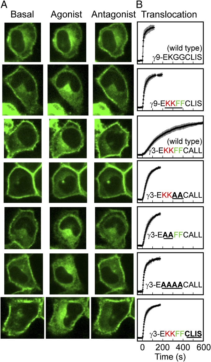
Fig. 6.
Hydrophobic and positively charged residues in the γ-subunit C-terminal domain determine rate of βγ translocation. (A) Confocal images of HeLa cells transfected with αo, β1, and a wild-type or mutant GFP-tagged γ-subunit. The specific γ-subunits are specified in the corresponding plots of the forward translocation kinetics in B. Data are plotted as the mean (±SD) from at least n = 14 cells and three independent experiments. Agonist addition corresponds to t = 0 s.

