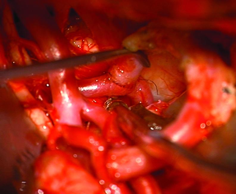
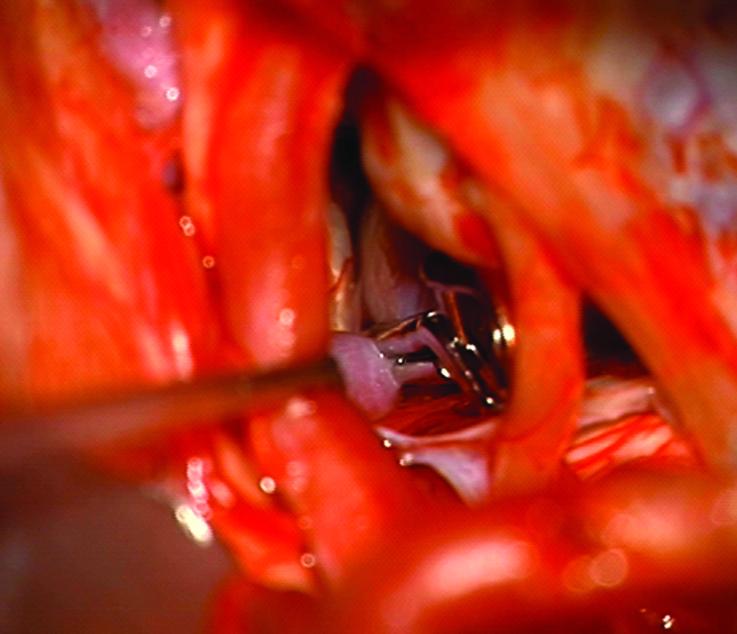
Figure 3.
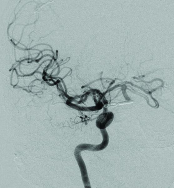
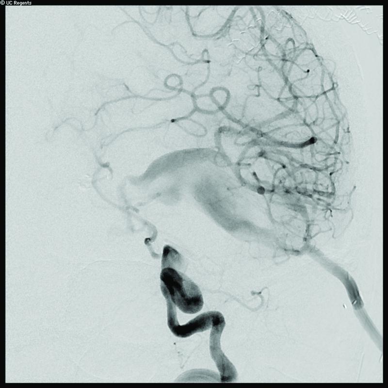
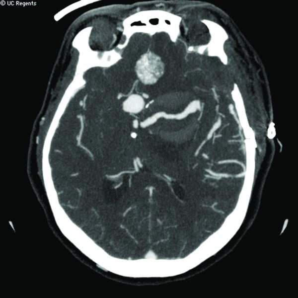
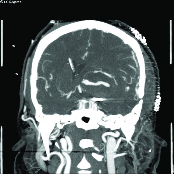
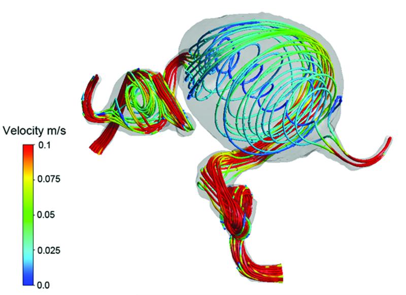
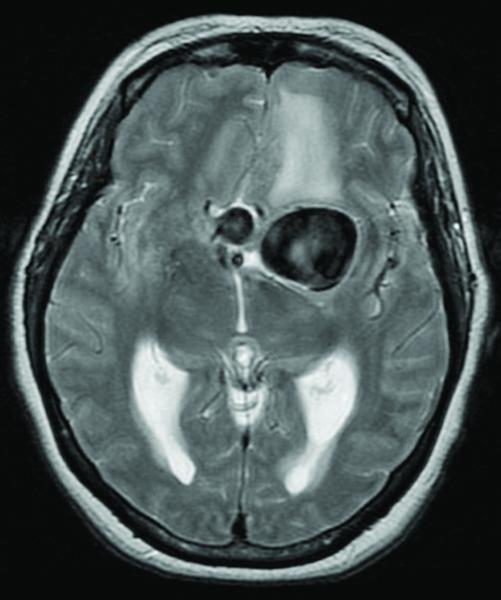
Case 15. (A) Axial T2-weighted MR imaging revealed a giant left ICA bifurcation aneurysm and a large anterior communicating artery aneurysm in this 51 year-old woman. (B) 3D reconstructed angiogram (left ICA injection) demonstrated its dolichoectatic morphology. An end-to-side anastomosis between radial artery and the efferent MCA was part of an ECA-MCA bypass. (C) Supraclinoid ICA was occluded with a clip as it entered the aneurysm, distal to PCoA. Indocyanine green videoangiography demonstrated patency of the bypass graft, filling of distal MCA branches, filling of the supraclinoid ICA up to the clip, and faint flow of dye within the aneurysm. Postoperative CT angiography showed a thin layer of new intra-aneurysmal thrombus anteriorly, posteriorly, and inferiorly on (D) axial and (E) coronal views. (F) Subsequent digital subtraction angiography demonstrated bypass patency and progressive intraluminal thrombosis (left ICA injection, anteroposterior view). CTA on postoperative day 5 revealed further intraluminal thrombosis with two serpentine channels connecting the bypass with the A1 segment on the opposite side of the aneurysm, as seen on (G) axial and (H) coronal views. Post-surgical thrombosis occluded the anterior choroidal artery, and she suffered a capsular infarct. This case demonstrates that post-surgical aneurysm thrombosis after proximal clip occlusion can occlude small branch arteries.

