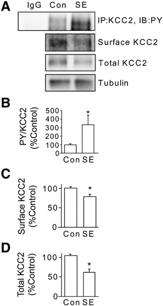
Fig. 7.
Induction of status epilepticus (SE) leads to tyrosine phosphorylation and degradation of KCC2. A. Adult FVB/N mice (8–12 weeks old) were injected with pilocarpine (330 mg/kg) to induce SE. Animals injected with saline were used as controls. Animals that developed SE for 1 h were sacrificed and whole brain 350 mM hippocampal slices were prepared. After incubation some slices were lyzed and KCC2 was immunoprecipitated and immunoblotted with KCC2 antibodies (upper panel). Slices were also labeled with biotin cell surface and total fractions were then immunoblotted with KCC2 antibodies or total fractions were immunoblotted with tubulin antibodies. Non-specific IgG was used as control for immunoprecipitation. These data were then used to calculate the ratio of PY/total KCC2 immunoreactivity (B), cell surface (C) and total levels (D) of KCC2. In each case data were normalized to control. [*Significantly different from control condition (p<0.01; value = mean ± SEM, Student's t-test, n=3 mice per group)].

