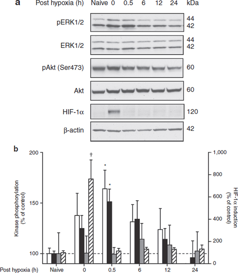
Figure 1.
Hypoxic preconditioning transiently activates extracellular-regulated kinase (ERK) in neonatal brain cortex. Postnatal day 6 mice were exposed to 45 min of global hypoxia (8% oxygen) and killed at various time points (0–24 h) after reoxygenation. Animals exposed to normoxia served as baseline controls (naive). Phosphorylation of ERK1 (white bars), ERK 2 (black bars), and Akt (gray bars), and hypoxia-inducible factor (HIF)-1α stabilization (hatched bars) were analyzed in cortical homogenates by western blotting as described in Methods. (a) Representative blots for a complete time course of one set of animals. (b) Bar graph shows a summary of the results from densitometric analysis of bands from n = 8 (phospho-ERK/ERK), n = 5 (pAkt/Akt), and n = 3 (HIF-1α) independent sets of animals. The level of phosphoprotein is expressed relative to total protein and to that of wild-type naive mice (=100%) ± SEM. *P < 0.05, †P < 0.001, by one-way ANOVA.

