
Figure 1.
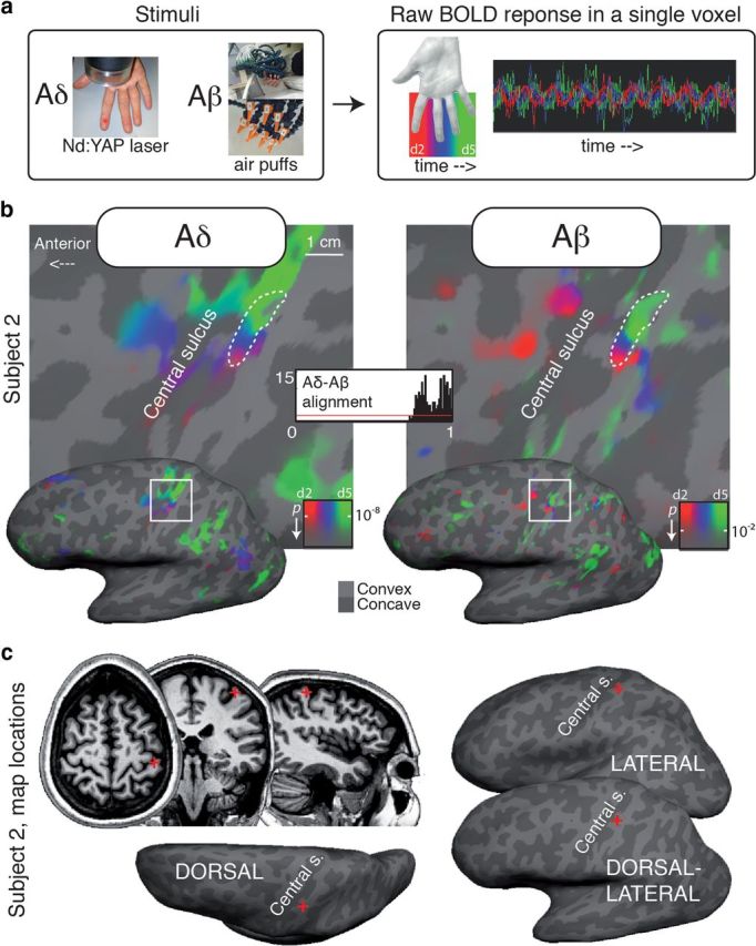
a, Phase-encoded protocol and raw BOLD response in one voxel. The color coding scheme used for Aδ (laser) and Aβ (air puffs) maps in Figures 1–3 is shown in a. b, Aligned Aδ (laser) and Aβ (air puffs) maps for a single subject (subject 2, dorsolateral view). Thick dashed white contours outline a region of interest defined as the connected surface patch of SI vertices with significant periodic response to both Aδ and Aβ stimulation. A similar alignment is evident for every color, representing stimulation to the digits (d2–d5). The alignment index histogram shows the distribution of agreement in phase angle for each surface vertex within the dashed contour (1 = max alignment). The red line indicates the distribution of the alignment index that would be expected if the two maps were completely uncorrelated. c, Location of the two maps (red cross) in an illustrative single subject.