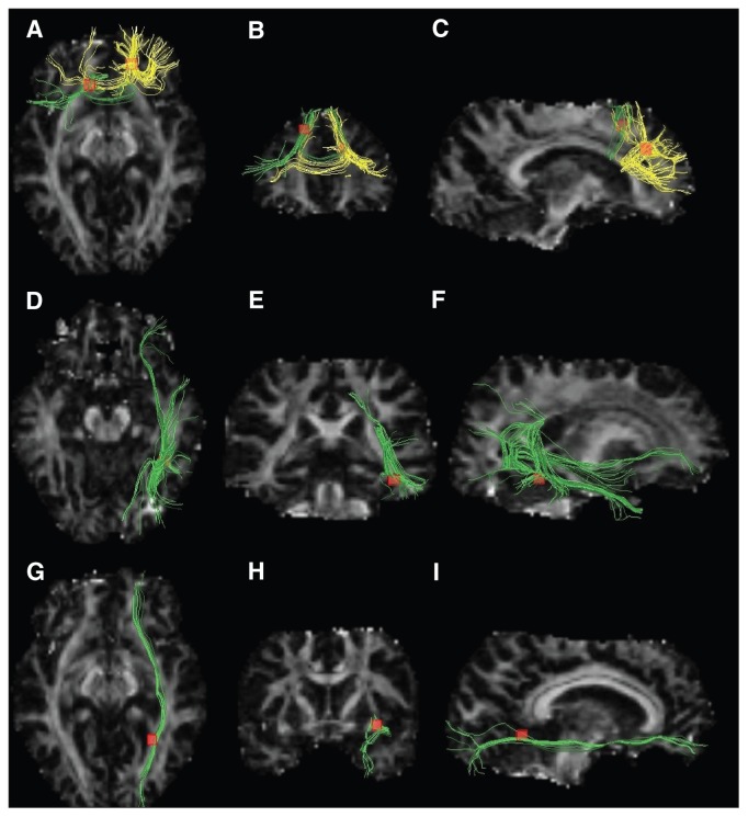
Fig. 3.
White mater diffusion tensor tracts traversing decreased fractional anisotropy clusters. Images on the left (A, D, G) are seen from above, medial images (B, E, H) are seen from behind and images on the right (C, F, I) are seen from the right. Fascicles traversing the right and left frontal white matter regions (yellow and green fibres, respectively, in the first row) were the genu and the body of the corpus callosum (A, B, C). Fascicles traversing the right fusiform gyrus white matter region were the right inferior longitudinal fasciculus, right interior fronto-occipital fasciculus and right posterior thalamic radiation (green fibres in the second row; D, E, F). Fascicles traversing the right occipital white matter region (yellow and green fibres, respectively, in the first row) were the right inferior fronto-occipital fasciculus (green tracts in the third row; G, H, I).

