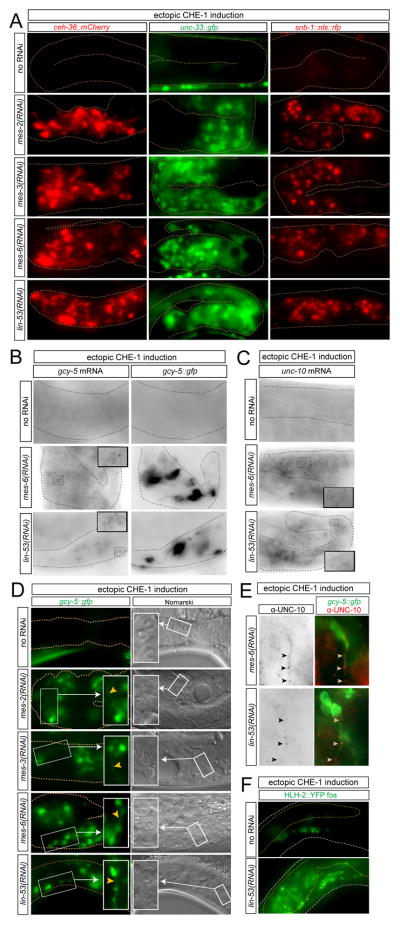
Fig. 2. Detailed characterization of germ cell-to-neuron conversion.
(A) The progeny of RNAi-treated animals were analyzed for the expression of several additional markers (ceh-36: otIs264; unc-33: otIs118; snb-1: otEx4445) after ~24 hours of heat-shock promoter-mediated che-1 induction at larval stages (otIs305 transgene). The penetrance of this phenotype ranged from 20% to 50% for the various markers (n = 30 to 60 for each marker, for each RNAi).
(B,C) Single molecule FISH shows induction of endogenous genes in converted germ cells. gcy-5 (B) and unc-10 (C) is induced in the germline of animals in which either lin-53 or the representative PRC2 complex component mes-6 was knocked down by RNAi and in which CHE-1 expression was ectopically induced. Individual mRNA molecules show up as individual black dots. In panel B, the gcy-5::gfp transgene (which only contains the gcy-5 promoter and will therefore not be picked up by the smFISH probes) allows to visualize individual, converted cells.
(D) Germ cells acquire a neuron-like morphology in terms of nuclear morphology (“fried egg” germ cell nuclei to “speckled” neuronal cell nuclei; right panels, including blow-up in white box) and axo/dendritic extensions (arrowheads; left panels, including blow-up in white box).
(E) Converted germ cells express the presynaptic protein UNC-10/Rim, which clusters along the length of a neuronal extension (arrowheads).
(F) Induction of the immature neuronal marker hlh-2, as assessed using a fosmid reporter transgene (otEx4720).

