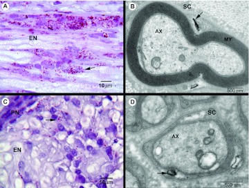
Fig. 2.
Inflammation,M. leprae infiltration and demyelination in infected armadillo PT nerve. (A) Longitudinal section showing a large number of acid-fast bacilli (AFB; arrow) in endoneurium (EN) of nerve. (B) Electron micrograph of a myelinated Schwann cell (SC) infected with M. leprae (arrow). AX, axon; MY, myelin sheath. (C) Cross-section of an infected nerve showing infiltration of M. leprae (arrow) in EN and perinurium (PN), as well as an infiltrate of mononuclear cells at the site of infection. (D) M. leprae (arrow) infecting non-myelinated Schwann cells in infected armadillo nerve.

