
Fig. 3.
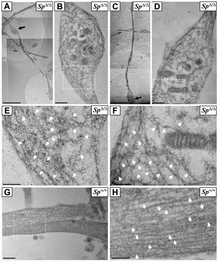
Ultrastructural analysis of SpΔ/Δ cortical neurons reveals a massive disorganization of the microtubule network within axonal swellings. (A–H) Ultrathin sections of DIV6 primary cultures of cortical neurons from SpΔ/Δ (A–F) and Sp+/+ mouse embryos (G–H). Images B and D are higher magnifications of axonal swellings indicated by arrows in images A and C, respectively. Images E, F and H are higher magnification of boxed regions in images B, D and G, respectively. Note the obvious disorganization and the tangled and bent aspect of microtubules within axonal swellings of SpΔ/Δ cortical neurons (B,D–F). This abnormal appearance of microtubules was never observed in Sp+/+ axons, in which microtubules are always organized in parallel arrays (G–H). White arrows indicate microtubules (E,F,H). Scale bars: 5 μm (A,C); 0.5 μm (B,D,G); 0.25 μm (E,F,H).