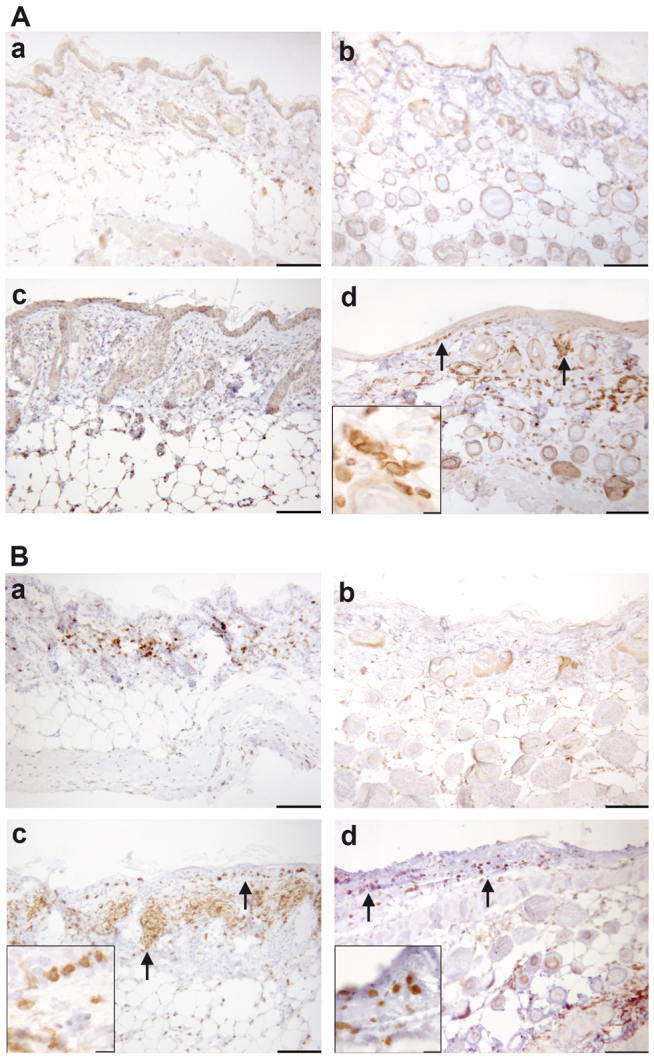
Fig. 2.

Upon challenge with oxazolone, human leukocytes (mainly consisting of T cells) infiltrate the dermis and epidermis. (A,B) Photomicrographs of immunohistochemically stained paraffin sections of the skin: (A) stained with anti-hCD45 antibody and (B) stained with anti-CD3 antibody. Skin samples for both A and B were taken from the following mice: BALB/c mouse challenged with ethanol (a); non-engrafted NOD-scid IL2Rγnull mouse (b); BALB/c mouse challenged with oxazolone (c); and engrafted NOD-scid IL2Rγnull mouse treated with oxazolone (d). Arrows indicate invaded human leukocytes. Scale bars: 100 μm; 10 μm (insets).
