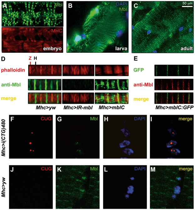
Fig. 6.
Sarcomeric distribution of Drosophila Mbl is reversibly altered by expression of CTG repeats. (A–M) Fluorescence images showing staining with anti-Mbl antibody. (A–C) Analysis of the endogenous Mbl expression (green) in the muscle throughout the different stages of development in wild-type individuals. In the embryonic somatic musculature, Mbl expression was restricted to the nucleus (A; compare with cytoplasmic signal of myosin heavy chain protein, MHC, shown in red below). (B) In the larval body wall muscles, Mbl was detected both in the nucleus (counterstained with DAPI in blue) and in cytoplasmic transversal bands. (C) In adults, the nuclear localization of Mbl was almost undetectable and the protein was preferentially detected in cytoplasmic transversal bands. (D) Double staining with anti-Mbl and phalloidin revealed that Mbl was localized in the sarcomeric H- and Z-bands. mbl silencing (Mhc-Gal4>UAS-IR-mbl), abolished Mbl signal in the bands, whereas overexpression of mblC (Mhc-Gal4>UAS-mblC), increased it mainly in the Z-bands. (E) GFP detection of a MblC:GFP fusion protein (green) coupled with an anti-Mbl antibody (red) confirmed the specificity of the signal in the Z-bands. However, MblC was not detected in the H-bands, suggesting a differential distribution of Mbl protein isoforms within the sarcomere. (F–M) Fluorescent confocal images comparing the subcellular distribution of i(CUG)480 RNA (fluorescent in situ hybridization using a CAG red-labeled probe; F,J) with Mbl protein (green; G,K; see merge in I,M) in control (J–M) and CTG-expressing flies (F–I). Nuclear signal of endogenous Mbl colocalized with CUG-RNA foci in CTG-expressing muscles. Nuclei were counterstained with DAPI (blue; H,L). (D–M) are confocal images using the 100× objective.

