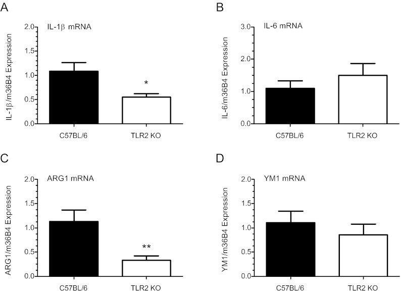
Fig. 7.
Amniotic fluid Mφs isolated from TLR-2-deficient mice exhibit decreased expression of M1 and M2 activation markers. F4/80+ AF Mφs isolated from TLR2−/− and WT B6 mice were isolated by FACSAria (BD Bioscience) at 18.5 dpc. Pooled Mφ mRNA (500 ng) from each timed-pregnant mouse was reverse transcribed and the expression of IL-1β and IL-6 was analyzed by qRT-PCR. Expression was normalized to m36B4 and calculated as fold change over WT control using the ΔΔCt method. Data indicate decreased levels of the classical activation (M1) marker IL-1β (A) (P < 0.05; n = 9) and comparable levels of IL-6 (B) (P = 0.4; n = 7) in TLR2−/− AF Mφ compared with WT. Analysis of alternative activation (M2) markers revealed significantly lower levels of ARG1 (C) mRNA (P < 0.001; n = 9) in TLR2−/− Mφs, whereas YM1 (D) (P = 0.4; n = 7) expression was similar to WT. Values are the mean ± sem. Statistically significant differences were analyzed by the Student's t test. *, P < 0.05, **, P < 0.001.

