Fig. 1.
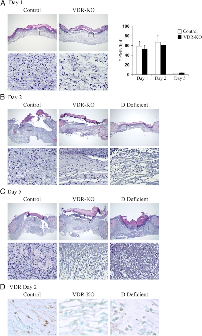
Abnormalities in the inflammatory response to cutaneous wounding are apparent in vdr−/− and vitamin D-deficient mice. Hematoxylin-and-eosin-stained sections from the center of wounds isolated 1 (A), 2 (B), or 5 (C) d after the injury from control, vdr−/− (VDR-KO), and vitamin D-deficient mice (D Deficient; housed in a UV free environment and fed a high calcium, high phosphorus, lactose supplemented diet lacking vitamin D metabolites). Boxes denote area magnified in bottom panel. Original magnification (top panel), ×4, (bottom panel), ×20. Data are based on at least two sections obtained per wound from wounds isolated from at least three mice per genotype or condition. Graph in A represents the number of polymorphonuclear cells/hpf. Data were manually quantified and averaged over 3 hpf from at least three mice per genotype/condition. Care was taken to exclude fibroblasts, macrophages, and endothelial cells and to avoid performing cell counts within the scale crust. D, Representative VDR IHC analyses 2 d after the injury for the VDR in the wound granulation tissue of control (left panel), vdr−/− (VDR-KO, middle panel), and vitamin D-deficient (D Def, right panel) mice. Original magnification, ×40. Data are based on at least two sections obtained per wound from wounds isolated from at least three mice per genotype or condition.

