
Fig. 3.
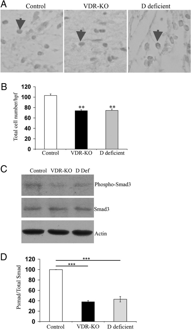
Activation of the TGF-β signaling pathway in response to cutaneous injury requires ligand-dependent actions of the VDR. A, Representative IHC analyses 2 d after the injury for nuclear pSmad3 in the wound granulation tissue of control (left panel), vdr−/− (middle panel), and vitamin D-deficient (right panel) mice. Arrows indicate examples of pSmad3 nuclear immunoreactivity. B, The total number of pSmad3-positive nuclei/hpf was quantified ±sem (n ≥ 3 mice). Statistical significance was determined using the Student's t test. **, P ≤ 0.001. Data are based on at least two sections obtained per wound from wounds isolated from at least three mice per genotype or condition. C, Total protein (20 μg) isolated from wounds of control (Ctrl), vdr−/− (VDR-KO), and vitamin D-deficient (D Def) mice 2 d after the injury was subjected to SDS-PAGE and immunoblotted for pSmad3 (top panel), total Smad3 (middle panel), and actin (bottom panel). D, The ratio of pSmad3 to total Smad3 was determined by the quantitation of signal intensity of the relevant bands. The ratio of pSmad3 to total Smad3 in the wounds of vitamin D-deficient and vdr−/− mice was normalized to that obtained from wounds of control mice. Statistical significance was determined using the student's test. ***, P ≤ 0.0005. Data represent those obtained from the wound protein lysates of four animals per genotype or condition.