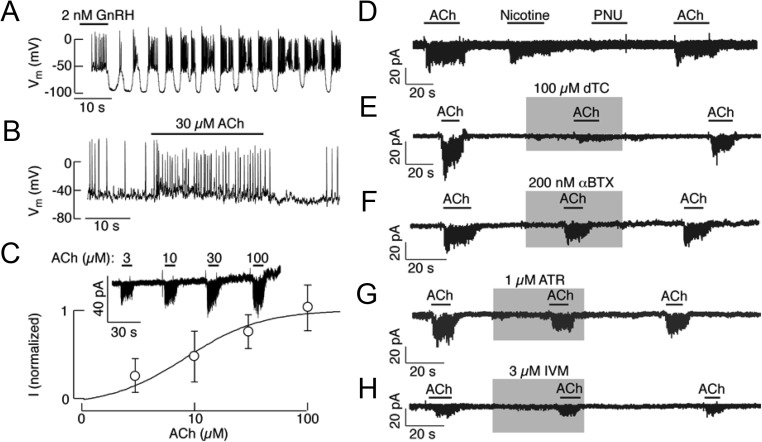
Fig. 7.
Electrophysiological characterization of nAChR in pituitary gonadotrophs. A and B, Current-clamp traces of GnRH (A) and ACh (B) induced electrical activity. Notice that GnRH-induced increase in the frequency of action potentials was periodically interrupted with transient hyperpolarization, whereas ACh application caused a sustained depolarization in this particular gonadotroph. C, Concentration-dependent effect of ACh on the amplitude of inward current in identified gonadotrophs. Top, Representative traces. Bottom, Mean ± sem values, with the estimated EC50 of 8.6 μm (n = 5). D, Stimulation of inward current by 100 μm ACh, 30 μm nicotine, but not by 1 μm PNU 282987. E, Inhibition of ACh-induced inward current by dTC. F–H, The lack of effect of αBTX, atropine (ATR), and ivermectin (IVM) on ACh-induced inward current in identified gonadotrophs. All traces were obtained using standard whole-cell patch-clamp recording. Gray areas and horizontal bars indicate duration of drug application.

