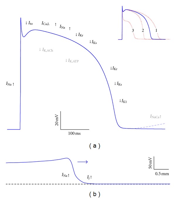
Figure 6.

Action potential formation and propagation in ventricular myocytes. (a) Diagram of an action potential in an isolated ventricular myocyte with individual ion currents that “push up” (inward currents, upward arrows) or “pull down” (outward currents, downward arrows) the action potential. I Ca,L: L-type calcium current; I K,ACh: acetylcholine-sensitive potassium current; I K,ATP: ATP-sensitive potassium current; I K1: inward rectifier potassium current; I Kr: rapid delayed rectifier potassium current; I Ks: slow delayed rectifier potassium current; I Na: fast sodium current; I NaCa: sodium-calcium exchange current; I to: transient outward potassium current. Grey symbols refer to currents that become important under special conditions (release of ACh, shortage of ATP and calcium overload for I K,ACh, I K,ATP, and I NaCa, resp.). Loss of function of “pull down” currents may result in action potential lengthening (inset, red dashed trace labeled “1”), whereas loss of function of “push up” currents may result in action potential shortening (inset, red dashed trace labeled “2”) and loss of the action potential dome (inset, red dashed trace labeled “3”). (b) Diagram of action potential propagation. The propagating action potential (horizontal arrow) brings neighboring cells to threshold through the I Na driven gap junctional current I j.
