Figure 1.
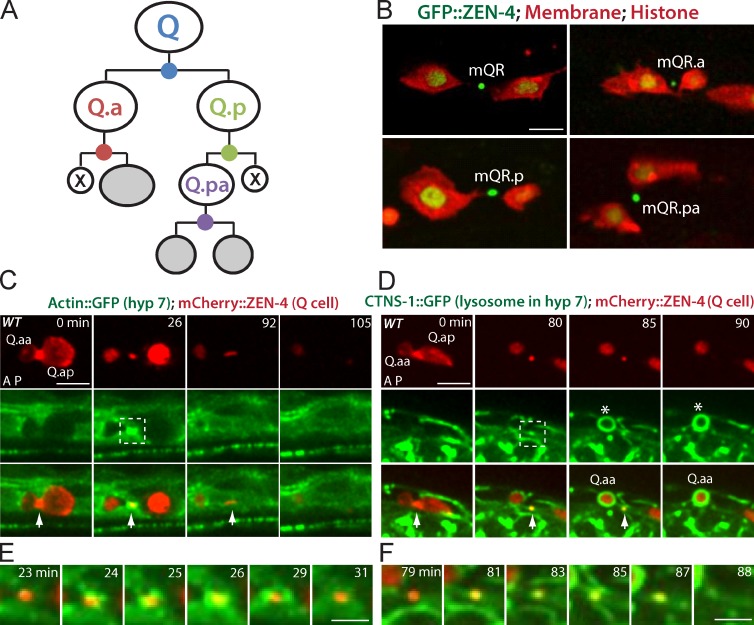
Q cell midbody is released after cell division and degraded by the hyp7 cell. (A) C. elegans Q cells are bilaterally symmetric, and both QR (right) and QL (left) cells undergo three rounds of asymmetric division to generate three different neurons (gray) and two apoptotic cells (X). Four divisions by the Q, Q.a, Q.p and Q.pa cell make four midbodies (dots in blue, red, green, and purple). (B) Midbody release in the QR lineage with midbodies (ZEN-4/MKLP1 and GFP::ZEN-4) shown and the plasma membrane (mCherry with a myristoylation signal) and histone (his-24::mCherry) shown. Midbody name is adjacent. (C) Still images of the birth, engulfment, and degradation of the Q cell midbody in WT. (D) Lysosome recruitment from the hyp7 cell to the Q cell midbody. (C and D) mCherry labels the Q cell midbody in C and D (top), and GFP marks actin cytoskeleton (C) or lysosome (CTNS-1; D) in the hyp7 cell (middle). Arrows show midbodies, and asterisks in D show the apoptotic cells. The cell name is adjacent. Anterior of the cell is to the left. Time in minutes is given on the top of top row. The birth of the Q cell midbody was the end of cytokinesis of the mother cell (0 min in C and D). The degradation was the last frame showing the Q cell midbody (105 min in C and 90 min in D). (E and F) Enlarged time-lapse views of the dotted boxes (26 min in C and 80 min in D) in which GFP-actin (E) and lysosome (F) from the hyp7 cell were recruited onto the midbody. Bars: (C and D) 5 µm; (E and F) 2.5 µm.

