Figure 2.
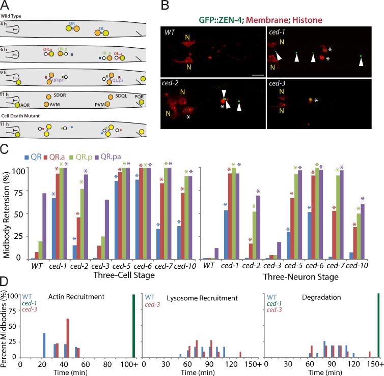
Apoptotic cell engulfment genes are required for midbody degradation. (A) Cartoon shows the Q cell midbody fate. At 4 h after hatching, the Q midbody was generated by QR or QL division; mQ.a or mQ.p and mQ.pa were generated at 6 or 9 h (three-cell stage), respectively. mQ, mQ.a, mQ.p, or mQ.pa was degraded at 6, 9, or 11 h (three-neuron stage). (bottom) In cell death mutants, the midbody failed to degrade. PVM, posterior ventral microtubule. (B) Fluorescence images of Q cell midbody degradation in WT and cell death mutants. Midbodies (ZEN-4/MKLP1 and GFP::ZEN-4), plasma membrane (mCherry with a myristoylation signal), and histone (his-24::mCherry) are shown. N shows neurons (anterior ventral microtubule [AVM] and SDQR). Arrowheads show persistent midbodies in ced-1 and ced-2 mutants. Asterisks show cell corpses in ced-1 and ced-2 mutants and the nonapoptotic cell in the ced-3 mutant. Bar, 5 µm. (C) Quantifications of midbody degradation in the QR cell lineage in WT and mutants at the three-cell (left) or thee-neuron (right) developmental stage. *, P < 0.01, χ2 test (mutant paired with WT). For each data point, n = 11–45 from a single experiment. (D) Quantifications of the time for Q cell midbody engulfment (actin recruitment in hyp7 cell), lysosome recruitment, and degradation in WT, ced-1, and ced-3 mutants. n = 11–26 from a single experiment. Statistic analysis is shown in Fig. S3.

