Fig. 1.
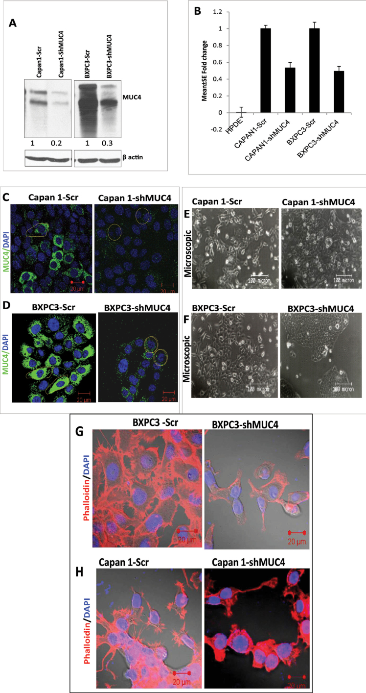
Strategy for shRNA-mediated silencing of oncogenic MUC4 in Capan1 and BxPC3 cells. (A) Analysis of MUC4 expression in BxPC3/Capan1-shMUC4, BxPC3/Capan1-Scr by immunoblotting shows significant inhibition in protein expression (70–80%), respectively. β-actin was used as a loading control. (B) Real-time PCR analysis using primers that specifically amplify the MUC4 gene. Capan1 and BxPC3 pooled population cells show a 50–60% decrease in the expression of MUC4 at the mRNA level compared with the control population. No amplification was observed in HPDE, which is negative for the MUC4 expression. (C and D). Confocal analysis showed a decreased expression of MUC4 on the cell membrane in Capan1 and BxPC3-shMUC4 cells compared with scramble cells. (E and F) A morphological comparison between the Capan1/BxPC3-shMUC4 and Capan1/BxPC3-Scr cells. The MUC4 knockdown Capan1 and BxPC3 cells grow in aggregates compared with the scramble cells. (G and H) The phalloidin-rhodamine (phalloidin-RITC) staining of actin–cytoskeleton. MUC4-expressing Capan1 and BxPC3-Scr cells revealed the presence of more microspikes, lamellopodia and filopodia-like cellular projections compared with the MUC4 knockdown Capan1/BxPC3 cells.

