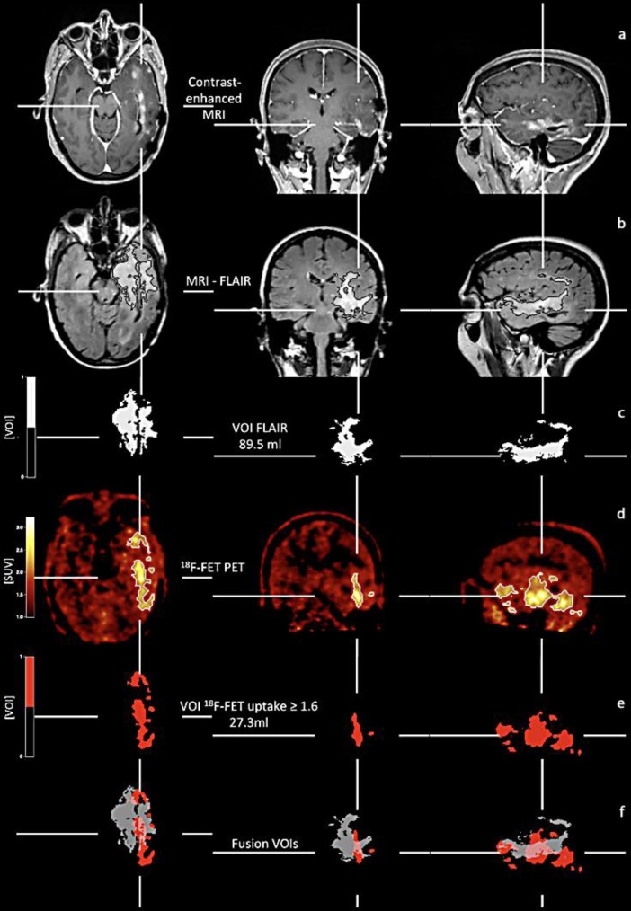
Fig. 1.
Coregistered MR, 18F-FET PET, and superimposed images of the metabolically active tumor volume (red) at an 18F-FET uptake index threshold of =1.6 (red) and volume of FLAIR hyperintensity (white). FLAIR hyperintensity is not always located exclusively within the subareas of the tumor positive on 18F-FET PET. On superimposed pictures (bottom), FLAIR hyperintensity (grey) is eccentric and located partially outside the metabolically active tumor volume (red). VOI = Volume of interest; SUV = standardized uptake value.

