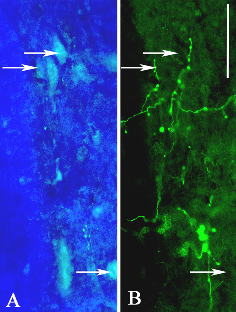
Figure 4.
High magnification, dual immunofluorescent images at the mid-thoracic spinal leel two weeks after T4 transection demonstrate close proximity of (A) FluoroGold-labeled sympathetic preganglionic neurons (arrows) in the intermediolateral cell column and (B) biotinylated dextran amine (BDA)-labeled fibers originating from lumbosacral projection interneurons. Scale bar = 50 µm.

