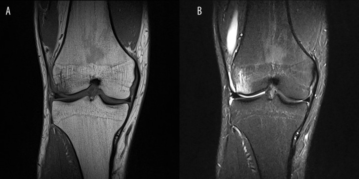
Figure 3.
(A) Coronal T1-wieghted images of the knee joint in a 30yo patient after knee injury. Decreased signal intensity is visible in the marrow cavity (signal intensity of the muscles is lower than of marrow cavity), reaching but not extending beyond the growth plate. Patient is an amateur long-distance runner, with weekly routine over 70 km. (B) PD fat saturation images show decreased signal intensity of yellow bone marrow. The image also reveals the presence of bone marrow edema on the lateral femoral epicondyle. Signal intensity of edematous bone marrow is higher than in metaphyseal areas of reconversion.

