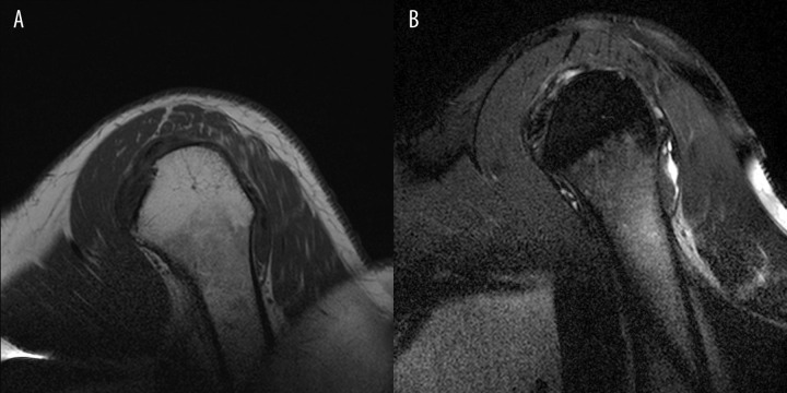
Figure 4A, B.
(A) Sagital oblique T1-weighted images of a 45 yo patient with shoulder pain shows areas of lower signal intensity in the metaphyseal area corresponding to bone marrow reconversion, which do not extend beyond the growth plate. Clinical data: smoking habit. (B) Sagital oblique PD fat saturation images in the same patient.

