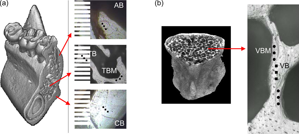
Fig. 1.
Descriptive regions for nanoindentation in 3D micro-CT images and under an indenter microscope; (a) Four mandibular regions (AB, alveolar bone (black dots); CB, cortical basal bone (black squares); TBM, marginal region of trabecular bone (black dots); TB, inner region of trabecular bone (black squares)) and (b) two vertebral regions (VBM, marginal region of vertebral bone (black dots) and VB, inner region of vertebral bone (black squares)).

