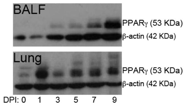
Figure 1.

Peroxisome proliferator-activated receptor (PPAR)γ protein expression during influenza virus infection in cells obtained by bronchoalveolar lavage fluid (BALF) (top), and lung (bottom). WT mice were infected with 5 × 10⁴ tissue culture infectious dose 50 (TCID50) of influenza A/Udorn (H3N2) and sacrificed 1,3,5,7 or 9 days post-challenge. Non-infected mice (0) were used to assess baseline PPAR γ expression at day 0. Influenza virus infection upregulated PPAR γ protein, reaching highest expression at day one post-infection in whole lung and on day 9 post-infection in cells obtained from the bronchoalveolar space. Results from BALF correspond to cells pooled from at least 3 mice at each time point. Results from lung correspond to a sample obtained from one mouse at each time point and the picture is representative of 4 replicates with identical results.
