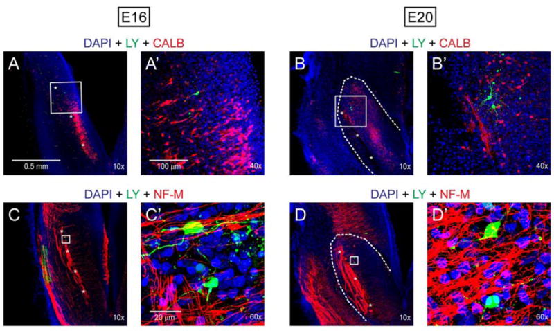
Fig. 1.

Post-recording immunohistochemistry in E16 and E20 brainstem slices. Recorded neurons (green) were intracellularly labeled by Lucifer yellow (LY). A, A’, B and B’: Recorded neurons were located in a cluster of calbindin (CALB)-immunoreactive cells (red) surrounding ST (indicated by asterisks). C, C’, D and D’: ST and the fibers extending medially to presumptive NST were neurofilament (NF-M)- immunoreactive (red). Recorded neurons were located in a meshwork of the NF-M-immunoreactive fibers. Blue color in all images was derived from nuclear staining with DAPI.
