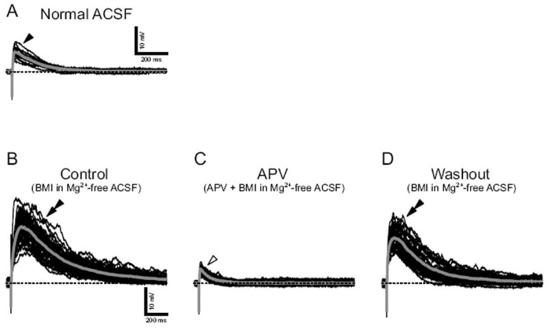
Fig. 7.

NMDA receptor-mediated excitatory postsynaptic potentials (EPSPs) evoked by current stimuli to ST in an E20 neuron. A: EPSPs in normal ACSF (filled arrowhead). B: The EPSPs enlarged in Mg2+-free ACSF (double arrowhead). C: The enlarged EPSPs were suppressed by a NMDA-type receptor antagonist APV (open arrowhead). D: The enlarged EPSPs reappeared after the washout of APV (double arrowhead). EPSPs were evoked by current stimuli repeated at 0.1 Hz. Baseline Vm was maintained at -70 mV (dash lines) with constant DC current.
