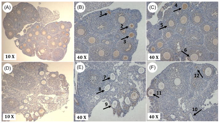
Figure 3. Lin28a protein levels decrease prior to the onset of puberty in ovarian tissue.
Immunohistochemical staining of Lin28A protein in 20 day (A–C) and 30 day (D–F) ovarian tissue. Lin28a protein expression is identified by diaminobenzidine staining. In 20-day-old tissue, Lin28a is detected in early developing follicles (arrow 1), early secondary follicles (arrows 2, 3, and 4), late secondary follicles and granulosa cells surrounding secondary follicles (arrow 5). In 30-day-old tissue, Lin28a protein is less evident, due to the abundance of atretic follicles (arrows 7, 8, 9 and 10). Lin28a still persists in the granulosa cells and oocytes of secondary follicles (arrow 11), and in early follicles (arrow 12). This pattern of Lin28a protein distribution was consistent in the two biological replicates that were examined

