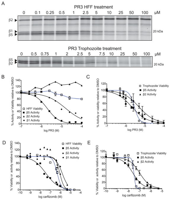
Figure 5.
PR3 is non-toxic to host cells due to partial inhibition of the human proteasome. A) MV151 labeling of intact HFF (top) and P. falciparum trophozoites (bottom) cells treated with PR3. Quantification of each labeled subunit (indicated by arrow) is shown below. B–E) Comparison of viability (blue line) versus inhibition of the catalytic proteasome subunit activities (black lines) after 1 hr incubation of HFF (B, D) or trophozoites (C, E) with PR3 (B–C) or carfilzomib (D–E). Proteasome activity was determined by competition of MV151 labeling from the gels shown in (A) and supplementary Figure S5. The effect of carfilzomib and PR3 in live trophozoites are obtained from 3 independent experiments; error bars represent S.E.M for each drug concentration from triplicates. (See also Figure S5).

