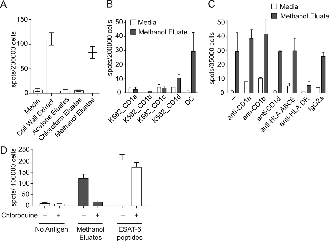
Figure 4.
Restricting element for presentation of antigens in cell wall extract. (A) Cell wall extract from M. tuberculosis and sub-fractions (10 µg/ml) were co-incubated overnight with PBMC and DCs from a subject with latent tuberculosis infection. (B) Sorted T-cells were co-incubated overnight with methanol eluates and either DCs or K562 cells transfected to express CD1a, CD1b, CD1c, or CD1d. (C) Sorted T-cells were co-incubated overnight with DCs, methanol eluates, and 10 µg/ml blocking antibody against CD1a (OKT6), CD1b (BCD1b.3), CD1d (CD1d42), MHC Class I and HLA-E (W6/32), HLA-DR (L243), or isotype control (IgG2a). (D) DCs were pre-incubated with 25 µM chloroquine for 15 minutes. These were co-incubated overnight with sorted T-cells in the continuous presence of 25 µM chloroquine and either 10 µg/ml methanol eluates or 10 µg/ml ESAT-6 peptides. In all cases, IFN-γ production was assessed by ELISPOT.

