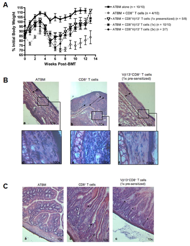
FIGURE 6. CBA recipients of tumor-presensitized B10.BR CD8+Vβ13+ T cells exhibit minimal histological evidence of GVHD.

CBA mice were exposed to lethal irradiation (11 Gy, split dose) and transplanted with 2×10⁶ ATBM cells alone or in combination with either CD8+ T cells (1×10⁷) or CD8+Vβ13+ T cells (1x, 8.34×10⁵ or 3x, 2.5×10⁶) from MMC6 presensitized or naïve B10.BR donor mice, as indicated. A, The mean ± SE % initial body weight of surviving mice in each group was derived relative to the mean weight of the group on d 0. n = # mice surviving at termination of experiment/ # mice at initiation of experiment. B, Comparison of the dermis and epidermis of the ear between mice transplanted with ATBM cells alone, CD8+ T cells, or CD8+Vβ13+ T cells (1x presensitized). H&E reveals normal and regular thickness of the epithelium (double arrows) of the ATBM [a] and CD8+Vβ13+ T cells (1x presensitized) specimens [c], while there is severe irregularity of the epidermal-dermal border (single arrows) in the CD8+ T cell sample [b] with evident hyperplasia of the epidermis. Likewise, the collagen layer in the dermis (double arrow) is regular and well arranged in the ATBM and CD8+Vβ13+ T cell sample [a&c] and has low cellularity [d&f]. In contrasts, the dermis in the CD8+ T cell sample exhibits increased thickness and vast cellularity (double arrow) [e]. C, comparison of the epithelium of the small intestine between mice transplanted with ATBM cells alone, CD8+ T cells, or CD8+Vβ13+ T cells (1x presensitized). H&E reveals the integrity of the epithelium in the ATBM specimen [a] while there is loss of the architecture of the villi and crypts in the CD8+ T cell sample (arrows) [b] compared to the more defined epithelial structure observed in the CD8+Vβ13+ T cell (1x presensitized) sample (arrows) [c].
