
Fig. 1.
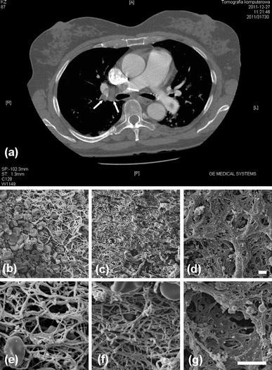
Computed tomography and scanning electron microscopic (SEM) images. Contrast-enhanced computed tomography showing a thrombus at the trifurcation of right pulmonary artery (a). Representative SEM images of the removed parts of the thrombus from the right atrium (b and e), lobar pulmonary artery (c and f) and segmental artery (d and g). b–d magnification, ×1,000; e–g magnification, ×3,500. Scale bar 5 μm