
Figure 4.
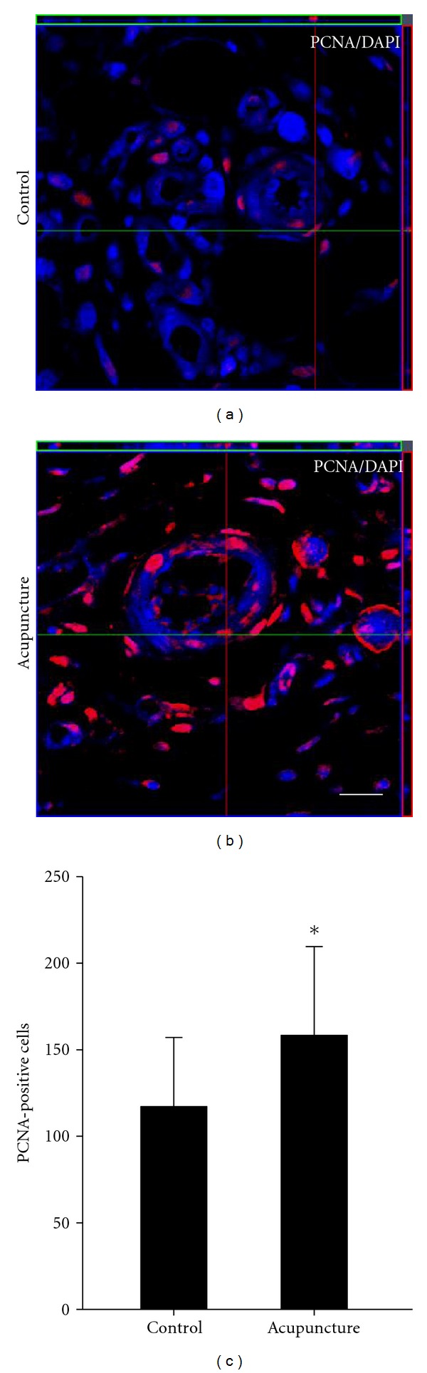
Quantitative analysis of PCNA-labeled cells in the wound area. Cell proliferation was measured by immunostaining using anti-PCNA antibody. At 7 days after-wounding, PCNA-labeled cells were present in wound area (a-b). PCNA positive cells were counted. In acupuncture-treated group, proliferation of endogenous cell was significantly increased compared to the control group (c). Data are expressed as mean ± SD, *P < 0.05, Scale bars denote, 50 μm.