
Figure 5.
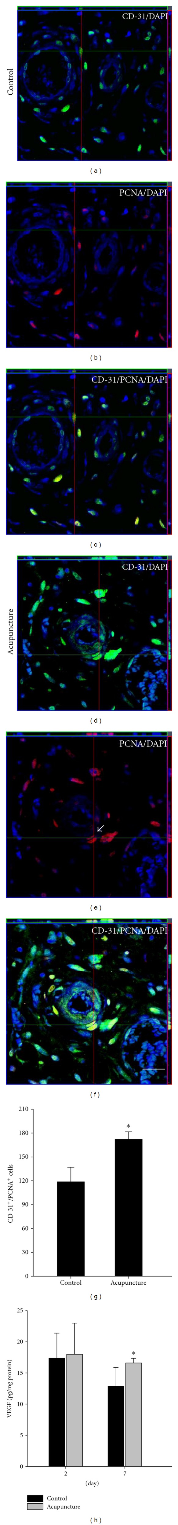
Quantitative analysis of angiogenesis by acupuncture in wound area. Histological analysis was shown into PCNA and CD-31 staining at 7 days after wound (a–f). The numbers of PCNA/CD-31-labeled cells were quantified present in wound area (g). At 7 days after-wounding, the expression of VEGF was significantly increased compared to the control group (h). Data are expressed as mean ± SD, *P < 0.05, Scale bars denote, 50 μm.