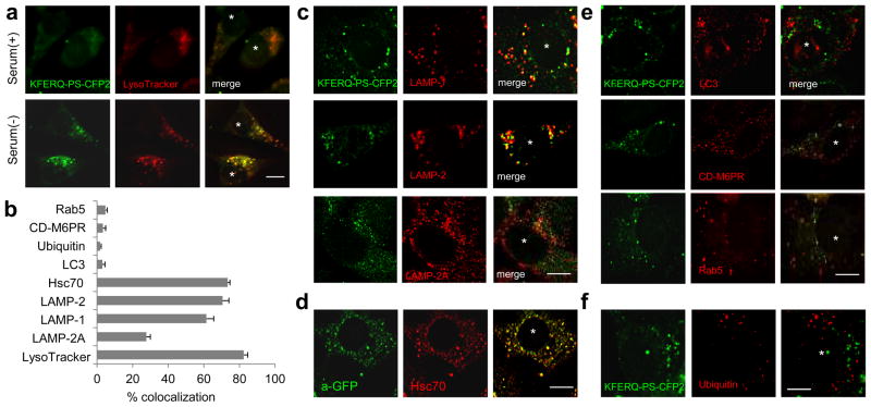
Figure 3. KFERQ-PS-CFP2 colocalizes with lysosomes after induction of CMA.
(a) Localization of photoconverted KFERQ-PS-CFP2 (green) in LysoTracker-stained compartments (red) in mouse fibroblasts maintained in the presence or absence of serum. (b) Percentage of colocalization of KFERQ-PS-CFP2 with the indicated proteins. Values are mean + SE of 3 different experiments with >50 cells counted per experiment. (c–f) Co-localization of photoconverted KFERQ-PS-CFP2 (green) with LAMP-1, LAMP-2 or LAMP-2A (b), LC3, CD-M6PR, Rab5 (d) and ubiquitin (e) in mouse fibroblasts maintained in serum-free media. d. Immunofluorescence with antibodies that recognize PS-CFP2 (green) and hsc70 (red) in cells maintained in the absence of serum and fixed with methanol to eliminate the soluble cytosolic fraction. Scale bars: 5μm

