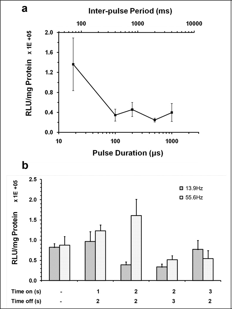
Figure 6. Effect of increased quiescent duration between US pulses on gene transfer efficiency.
(a) Effect of acoustic pulse durations on gene delivery. US exposure were performed on medial liver lobe for 90 sec, the first 45 s of which was coincident with injection of 150µg pGL4 plasmid and 15 Vol% Definity® mixture into medial lobe. The acoustic exposure conditions with various inter-pulse periods from 72 – 4000 ms (upper axis) and corresponding pulse durations (lower axis) from 18~1000 µs were 1.1 MHz, 2.7 MPa peak negative pressure, 0.00025 Duty Factor. Luciferase expression was measured on the treated median lobe. (b) Effect of pulse-train US exposure on gene delivery. US exposure was performed using either continuous bursts of 20 cycle pulses repeated at 13.9 or 55.6 Hz PRF for 90 s, or 1, 2 or 3 s exposures to bursts of pulsed US (on), interleafed by quiescent periods of 2 or 3 s (off). The detailed parameters were shown in Table 2 (lower panel). All groups were treated by 90 s US exposures with simultaneous 45 s injection of plasmid/MBs via the portal branch toward the hepatic median lobe. Luciferase expression was measured on the treated liver lobe. Error bars indicate SEM (n=6–7).

