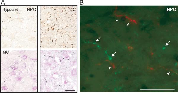
Figure 1.
Hypocretinergic and MCHergic fiber in the nucleus pontis oralis. A. Photomicrographs showing hypocretin-2 (upper photos) and MCH (lower photos) immunostained varicose fibers and terminals in the NPO and LC. These photomicrographs were obtained from 14 μm-thick sections that were processed with the ABC-DAB method. MCH immunostaining was enhanced by nickel and the sections were lightly counterstained with Pyronin-Y. B. Hypocretin-1 (in green) and MCH-containing (in red) fibers and terminals are intermingled in the NPO. Arrows and arrowheads indicate hypocretinergic and MCHergic fibers, respectively. Photomicrographs were obtained from sections that were processed for the detection of immunoflourescence. FITC (for Hcrt-1) and Rhodamine (for MCH) were used as fluorescent agents. This figure was produced by superimposing photomicrographs that were obtained using green and red filters. Calibration bars: A, 100 μm; B, 50 μm.

