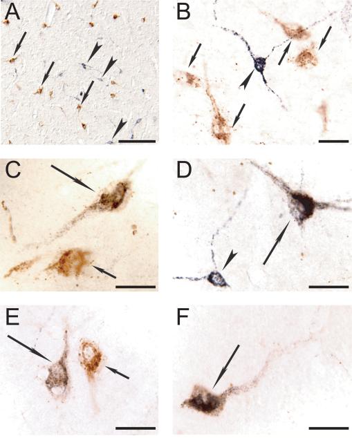
Figure 3.
Representative photomicrographs showing double immunolabeling for Hcrt and CTb in the postero-lateral hypothalamus. A. Photomicrograph of the perifornical region of the hypothalamus of PA8. Hcrt (stained in brown) and CTb (stained in black) labeled neurons are intermingled; examples are indicated by arrows and arrowheads, respectively. B to F. High magnification photomicrographs reveals double-labeled Hcrt+CTb+ neurons (long arrows) that are located in close relation with single labeled Hcrt+ neurons (small arrows) and single-labeled CTb+ neurons (arrowheads). Calibration bars: A, 150 μm; B to F, 30 μm.

