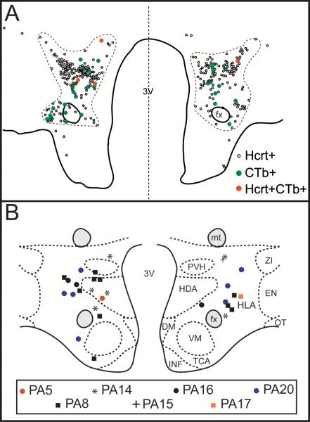
Figure 4.
Distribution of double-labeled Hcrt+CTb+ neurons. A. Camera lucida drawings of single-labeled Hcrt+ (small empty circles), single-labeled CTb+ neurons (green circles) and Hcrt+CTb+ neurons (red circles) at the tuberal level of the hypothalamus in cat PA14. Each dot represents one neuron. The dashed line demarcates the hypocretinergic field. B. Schematic drawing that depicts at the same hypothalamic level, the location of double-labeled Hcrt+CTb+ neurons that project to the NPO executive site for the generation of REM sleep. The neuronal distribution of 2 sections for each animal is presented. Different labels represent different cats; each label represents one neuron. DM, dorsomedial nucleus; EN, entopeduncular nucleus; fx, fornix; HDA, dorsal hypothalamic area; HLA, lateral hypothalamic area; INF, infundibular nucleus; mt, mamillothalamic tract; OT, optic tract; PVH, parvocellular nucleus; TCA, area of the tuber cinereum; VM, ventromedial nucleus; ZI, zona incerta; 3V, third ventricle.

