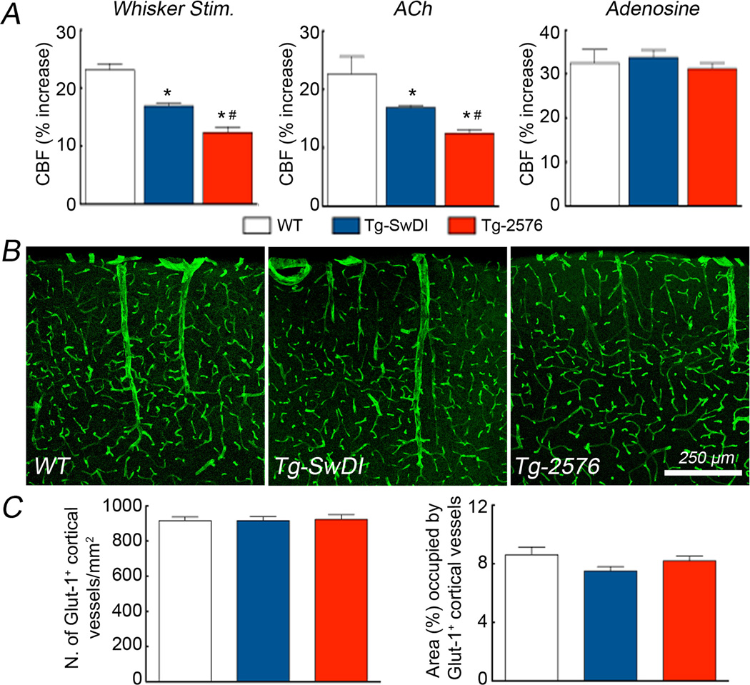
Figure 3.
Increases in CBF elicited by whisker stimulation (A), ACh (B) and Adenosine (C) in WT, Tg-SwDI and Tg-2576 mice (*p<0.05 from WT; # p<0.05 from WT and Tg-SwDI; Analysis of variance and Tukey’s test; n=5/group). Glut-1 immunoreactivity in the somatosensory cortex of WT (D), Tg-SwDI (E) and Tg-2576 mice (F). Number of vascular profiles (G) and % area occupied by blood vessels (H) do not differ among the groups (p>0.05; n=4–5/group).

