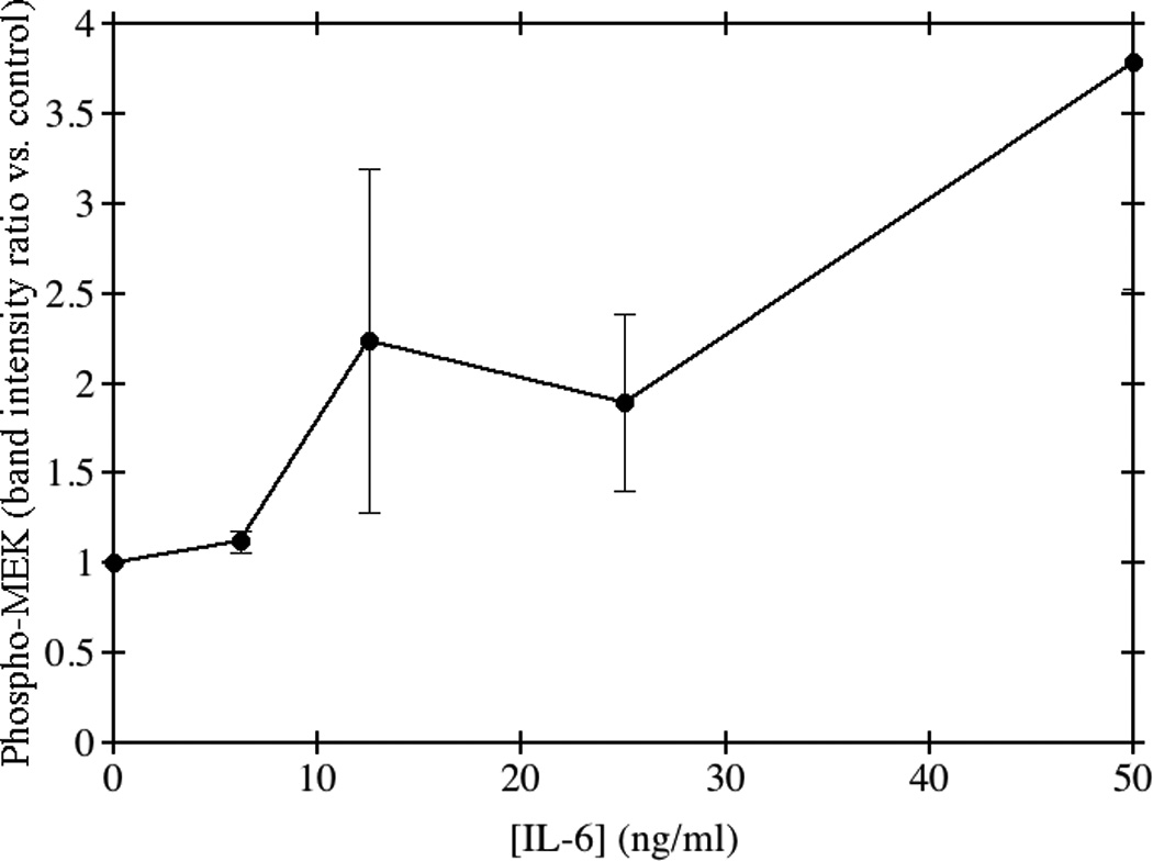
Fig. 6. Phosphorylation of MEK in response to IL-6.

HepG2 cells were grown to confluence and incubated with medium containing the indicated concentrations of IL-6 for 24 hours. Total cellular protein was separated using 10% SDS-PAGE and transferred to nitrocellulose membranes. The membranes were probed using a rabbit anti-human primary antibody to phospho MEK1/2 Ser217/221. Goat anti-rabbit HRP-conjugated secondary antibody was used and membranes were imaged by chemiluminescence. The average of triplicate experiments is shown ± SE.
