Figure 2.

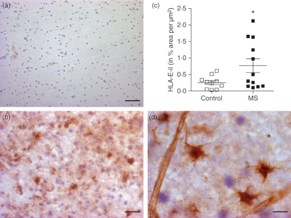
Increased HLA-E protein expression in multiple sclerosis (MS) white matter lesion. Little HLA-E immunolabelling (HLA-il) was detectable in white matter from control tissues (a) compared with MS white matter lesions (b). Staining was mostly detected on endothelial cells and astrocytes (d). (c) Levels were significantly up-regulated (P = 0.018; one-tailed) in MS tissue (0.767 ± 0.21) compared with controls (0.2541 ± 0.062). Scale bar = 30 μm for a; 20 μm for b, 10 μm for d.
