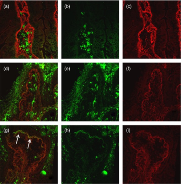
Fig. 1.

Duodenal mucosa from potential coeliac disease (CD) patient negative for mucosal deposits of immunoglobulin (Ig)A anti-transglutaminase 2 (TG2) before (a–c) and after 24 h culture with medium alone (d–f). Mucosal deposits of IgA anti-TG2 (in yellow) is lightly visible after 24-h culture with P31–43 (arrows) (g–i). IgA secreted by plasma cells are visible in green (b,e,h); TG2 with a subepithelial localization is shown in red (c,f,i).
