
Figure 4. Iodide decreases NIS mRNA levels in enterocytes.
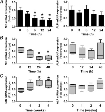
A, IEC-6 cell cultures were incubated with 100 μm I− for different periods of time. Quantitative PCR analysis was performed to quantify NIS and alkaline phosphatase (ALP) mRNA levels relative to those of β-actin. The expression level of untreated cells was set to 1. Values are indicated as fold change relative to the mRNA levels of untreated cells. *P < 0.01 vs. control (t= 0; ANOVA and Newman–Keuls test). B, male Sprague–Dawley rats (n= 6 per group) were treated with 0.05% I− in drinking water for the indicated periods of time. After treatment, villus tip small intestinal absorptive cells were isolated and qPCR analysis was performed to quantify mRNA levels relative to those of the loading control, β-actin. Data are indicated as the fold change relative to the mRNA levels of control animals (set as 1.0) and presented as box plots. *P < 0.05 vs. control (t= 0) (Kruskal–Wallis and Dunn's tests). C, male C57BL/6 mice (n= 5 per group) were subjected to an I−-deficient diet for 1–4 weeks. Quantitative mRNA analysis was performed as in B. *P < 0.05 vs. control (t= 0; Kruskal–Wallis and Dunn's tests).