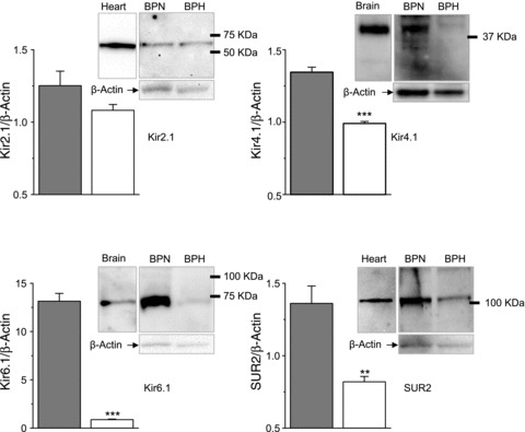
Figure 3. Expression of Kir2.1, Kir4.1, Kir6.1 and SUR2 proteins in BPN and BPH mesenteric VSMCs.

Representative immunoblots of mesenteric VSMC lysates from BPN and BPH arteries showing a band of the expected size for Kir2.1, Kir4.1, Kir6.1 and SUR2 as indicated. Loading control (β-actin) of the same membranes is also shown, as well as positive controls for the antibodies (brain lysates for Kir4.1 and Kir6.1 and heart homogenates for Kir2.1 and SUR2). Quantification of these data (bars plots) was obtained via densitometric analysis of 3–5 immunoblots for each antibody, normalized to their corresponding β-actin signals. **P < 0.01, ***P < 0.001.
