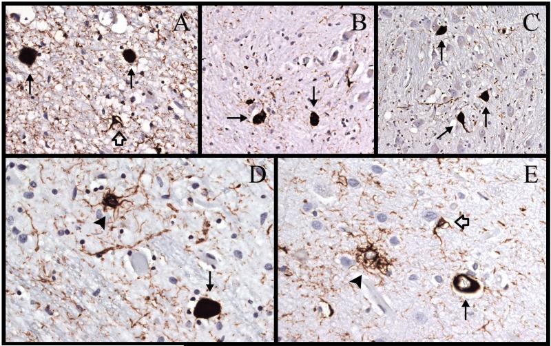
Figure 3.
Immunostaining for hyperphosphorylated tau (AT8) in case 3. (A) There are 2 neurofibrillary tangles ([NFTs], arrows) and a glial cytoplasmic inclusion (GCI) (open arrow) in the subthalamic nucleus. (B) There are 2 NFTs (arrows) and scattered neuropil threads in the dorsal arm of the inferior olivary nucleus. (C) There are 3 NFTs (arrows) in the pontine nuclei. (D) There is an NFT (arrow) and a tufted astrocyte (arrowhead) in the red nucleus. (E) There is an NFT (arrows), a tufted astrocyte (arrowhead), and a GCI (open arrow) in the superior parietal lobe. Magnifications: A, D, E, 400x; B, C, 200x.

