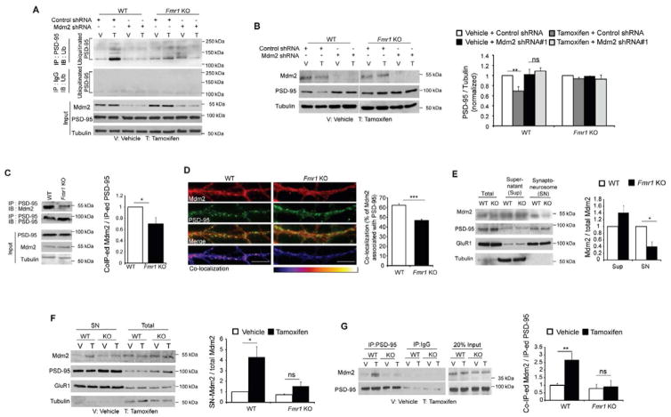
Figure 6. Mdm2 and Mdm2-mediated degradation of PSD-95 are dysregulated in Fmr1 KO neurons.
(A) Western blots of Ubiquitin after immunoprecipitation with PSD-95 (top) or IgG (second panel from the top). Protein samples were from dissociated WT or Fmr1 KO cortical neurons transfected with either control shRNA or Mdm2 shRNA together with MEF2-VP16ERtm. Treatment was as indicated and input was shown on the bottom. (B) Western blots of PSD-95, Mdm2 and Tubulin from dissociated WT or Fmr1 KO cortical neurons transfected with either control shRNA or Mdm2 shRNA together with MEF2-VP16ERtm. Quantification is shown on the right. (n = 3 cultures). (C) Western blots of Mdm2 immunoprecipitated with PSD-95 from WT or Fmr1 KO hippocampi. (n = 4 mice per genotype) (D) Immunohistochemistry of dendritic Mdm2 with PSD-95 from dissociated WT or Fmr1 KO hippocampal neurons. Co-localization images are shown on the bottom. Scale bars: 5 μm. Quantification of co-localization is shown on the right. (n = 21 cells) (E) Western blots of Mdm2, PSD-95, GluR1 and Tubulin in synaptoneurosomes prepared from WT or Fmr1 KO hippocampi. Quantification of Mdm2 in supernatant or synaptoneurosome is shown on the right. (n = 4 mice per genotype) (F) Western blots of Mdm2, PSD-95, GluR1 and Tubulin after synaptoneurosome preparation of dissociated WT or Fmr1 KO cortical neurons transfected with MEF2-VP16ERTm. Quantification of Mdm2 in synaptoneurosome is shown on the right. (n = 3 cultures) (G) Western blots of Mdm2 immunoprecipitated with PSD-95 from dissociated WT or Fmr1 KO cortical neurons transfected with MEF2-VP16ERtm. Quantification is shown on the right. (n = 3 cultures). In all figures, error bars represent SEM. (*p < 0.05, **p < 0.01, ***p < 0.001) (Experiments in A, F and G were done in the presence of MG132) See also Figure S6.

