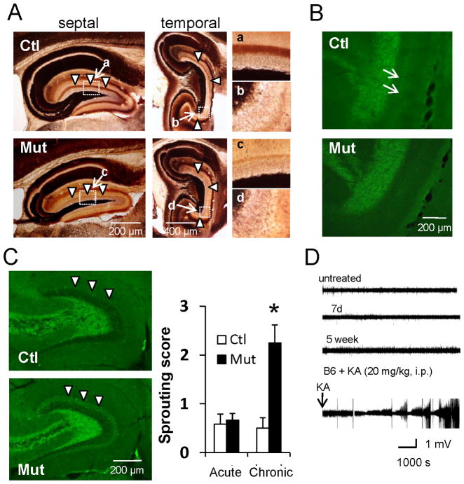
Figure 6. Functional changes after mossy cell loss.
(A) Timm staining of septal and temporal hippocampi during the chronic phase after mossy cell loss. Mutants show no mossy fiber sprouting and mossy cell axon terminal band-like staining in IML (arrowheads) almost disappears.
(B) ZnT3-IR in mutants shows no mossy fiber sprouting and loss of mossy cell axon terminal band-like staining in IML (see arrows in control).
(C) (Left) Hippocampal GAD67-IR during chronic phase. (Right) The sprouting score (modified Tauck and Nadler method) is higher in mutant IML than in controls (arrowheads). Mann-Whitney U-test (*p<0.05).
(D) Representative 2 hr traces of in vivo LFP recorded from mutant mouse before treatment (untreated) and 7 days (acute phase) and 5 weeks (chronic phase) after DT treatment. Epileptic LFP events following KA injection (20 mg/kg i.p.) to wild-type mouse are also shown. No obvious epileptiform discharges were observed following DT treatment.

