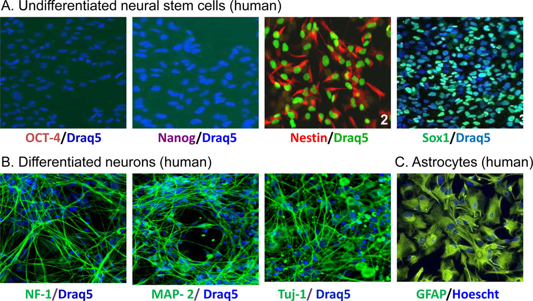
FIG. 1.
Fluorescence images of specific markers for stem cells and differentiated neurons. (A) Undifferentiated neural stem cells were positively stained for the NSC markers Nestin andSox1. (B) Differentiated neurons were positively stained for the neuronal marker NF-L, MAP2 & β-tubulin III. (C) Astrocytes were positively stained with GFAP. The colored words under each image represent the specific marker stained in the cells.

