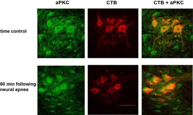
Figure 5.
aPKC levels in CTB-labeled phrenic motor neurons do not change 60 min following neural apnea. Representative photomicrographs depicting aPKC (green) and CTB (red) in the C4 ventral horn of time controls (top panel) or 60 min following neural apnea (iPMF; bottom panel). The merged image on right demonstrates colocalization of CTB(+) phrenic motor neurons and aPKC. Scale bar, 100 μm (at 20×).

